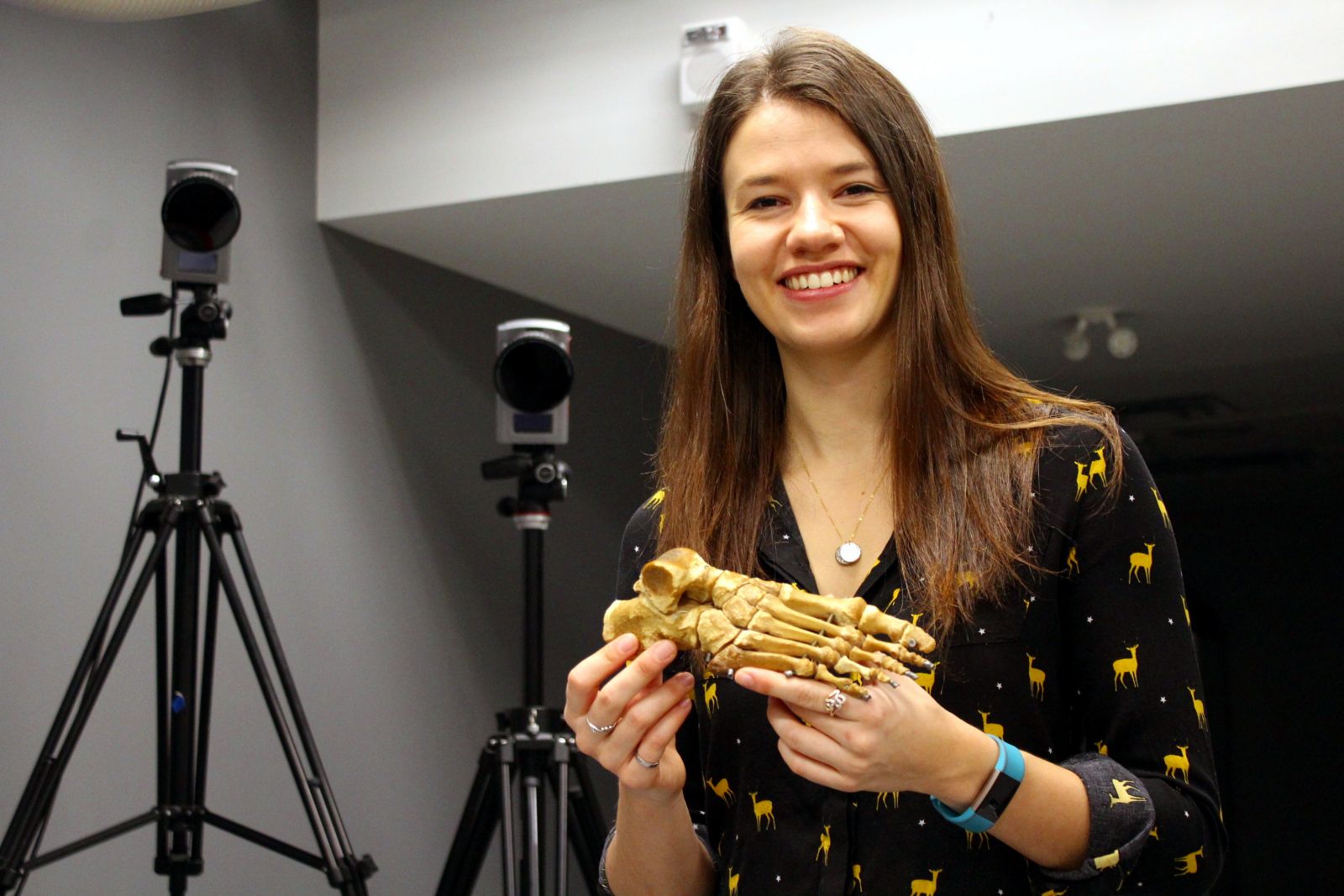
Thanks to advances in 3D x-ray imaging to measure complex foot function, ongoing research by Dr. Lauren Welte, a postdoctoral fellow at the Skeletal Observation Lab in the Department of Mechanical and Materials Engineering, is challenging nearly 70-year-old assumptions.
Published recently in the journal Proceedings of the Royal Society B, Welte’s discovery—in a collaborative study between several universities—is likely to have implications in shoe design and the treatment of foot problems.
Using high-speed x-ray to measure individual bone motion in the arch of the foot, researchers were able to model how the plantar fascia behaves in running. The discovery circled around the windlass mechanism.
“The plantar fascia is a ligament on the bottom of the foot that connects the heel to the toes and has the interesting characteristic of wrapping around the ball of the foot,” said Welte. “This wrapping means that when the toes lift up, it pulls the heel closer to the toes, lifting the arch, which is known as the windlass mechanism.”
Identified in 1954, the windlass mechanism states that the plantar fascia, which is an extensible tissue, doesn’t stretch enough to make a difference.
“We found that the toes are up when you land on the ground, and that the plantar fascia doesn’t stretch until later in stance, suggesting that your plantar fascia doesn’t manage the load when you hit the ground.”
If one’s toes are limited in motion, as in a form-fitted shoe, it could change the load applied to the arch tissues upon each step.
“We also found that the plantar fascia’s shortening, and energy return, is delayed at push-off,” Welte added. “This changes our fundamental understanding of how the foot works, which will likely have implications in shoe design and treatment of foot problems, among others.”
“Publishing her findings in a top-rated international journal is a testament to Lauren’s excellence as a researcher, the exceptional research training and laboratory facilities provided by Dr. Rainbow and his collaborators, and the comprehensive undergraduate education that Lauren received as a graduate of our biomechanical engineering option,” said Dr. Keith Pilkey, who heads the Department of Mechanical and Materials Engineering at Queen’s.
These discoveries follow previous work Dr. Welte has conducted regarding biomechanics and foot function, which was published in 2018. Now a postdoctoral fellow in the Department of Mechanical and Materials Engineering, she successfully defended her PhD thesis in 2020. This project was funded by the NSERC Discovery Grant, the NSERC Postgraduate Doctoral Scholarship and the Australian Research Council. Dr. Welte’s research was conducted under the supervision of Dr. Michael Rainbow.
“It was an absolute pleasure to work on this project with Lauren,” said Dr. Rainbow. “It really highlights the potential of dynamic x-ray imaging to provide new insights into human musculoskeletal function.”